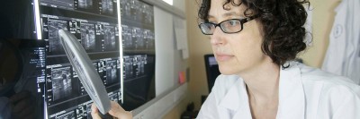
Radiologist Yolanda Bryce (right) and ultrasound supervisor Van Castor are part of a team that provides high-quality imaging services at MSK.
Physicians use imaging tests to help detect and diagnose disease, make appropriate treatment recommendations, and monitor your response to therapy.
Some imaging tests, like x-rays and computed tomography (CT), use radiation to capture images of the body. Others, like ultrasound and magnetic resonance imaging (MRI), do not. Because the various types of imaging tests provide different information, they’re not always interchangeable.
Your safety is one of our top concerns. If you must have a test that requires the use of radiation, our doctors take special care to reduce the radiation dose as much as possible without compromising the quality of the images needed for evaluation.
We will not order a test unless it is justified, meaning that the benefit of having the exam outweighs any potential risk. We’ll be sure to take the time to answer your questions and address any concerns you may have about a given imaging test. Our goal is to obtain the very best information possible so that we can make informed diagnostic and treatment recommendations and engage with you in shared decision making about your care.
Common Imaging Tests that Use Radiation
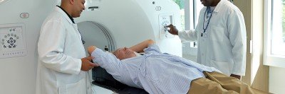
Computed Tomography (CT)
During a CT scan, a special machine uses x-rays to create a series of detailed, computerized pictures of areas inside the body taken from different angles. The three-dimensional images reveal abnormalities in both bone and soft tissues, including organs, muscles, and tumors. For some CT exams, a special material called a contrast dye is used to make the area of the body being studied easier to see.
A special CT scan that utilizes lower doses of radiation may be used as a screening tool for some individuals at high risk of developing lung cancer.
Nuclear Medicine Tests
Nuclear medicine tests use small amounts of radioactive material to diagnose disease. The radioactive substance is injected into the body, locates specific cells or tissues ― including cancer cells ― and binds to them. Images are made using a special machine (such as a PET scanner) that detects the radioactive substance, helping physicians to find cancer, see how far it has spread, or gauge how well a treatment is working.
Bone Scans
A bone scan is used to examine the bones for damage caused by cancer that either started there or that has spread from another part of the body. A small amount of radioactive material is injected into a vein. It travels through the bloodstream, collects in the bones, and is detected by a scanner, which creates images of the bones on a computer screen.

Positron Emission Tomography (PET)
During a PET scan, a small amount of radioactive glucose (sugar) is injected into a vein. A scanner then takes detailed, computerized pictures of areas inside the body where the glucose is being used. Because cancer cells often use more glucose than normal cells, the pictures can help physicians find cancer cells in the body.
In some cases, your doctor may recommend an integrated PET-CT scan. This combines images from PET and CT scans that have been performed at the same time using the same machine. Together, these two scans create a more complete picture of what is going on in the body than either test can offer alone.
Mammography
Mammography uses a low-dose x-ray system to create images of the breast that so doctors can detect cancer. Recent advances in technology have helped enhance the images produced. For example, digital mammography captures images of the breast that can be seen on a computer screen, and computer-aided detection (CAD) software can search digitized mammographic images for abnormal areas of the breast that require further analysis.
Breast tomosynthesis is a mammography system that creates a series of three-dimensional images of the breast that improves the physician’s ability to detect breast cancer and results in fewer patients having to undergo additional imaging.
X-ray
X-rays are the oldest and most frequently used form of medical imaging. This noninvasive test involves exposing an area to a small dose of radiation to create pictures of the inside of the body.
Common Imaging Tests that Do Not Use Radiation
Magnetic Resonance Imaging (MRI)
MRI uses radio waves and a powerful magnet linked to a computer to create detailed pictures of areas inside the body. These pictures can show the difference between normal and diseased tissue. MRI makes better images of organs and soft tissue than other scanning techniques, such as CT or x-ray.
High-quality images are assured only if you are able to remain perfectly still and hold your breath at specific times while the pictures are being recorded. MRI may take more time to perform than other imaging techniques, and some people ― particularly children or people with anxiety or a fear of enclosed spaces ― may find it difficult to lie still during imaging.
Metal and electronic objects can interfere with the magnetic field of the MRI unit. You should tell the technologist if you have medical or electronic devices or other metal objects in your body because they may pose a risk, depending on their nature and the strength of the MRI magnet.
Ultrasound
This safe and painless procedure produces pictures of the inside of the body using high-frequency sound waves. The sound waves are transmitted through a transducer, or probe, that is placed directly on the skin over the area that needs to be imaged. The sound waves are bounced off internal tissues or organs and form echo patterns that are shown on the screen of an ultrasound machine. Because the images, called sonograms, are captured in real time, they can show the structure and movement of the body’s internal organs, as well as blood flowing through blood vessels.